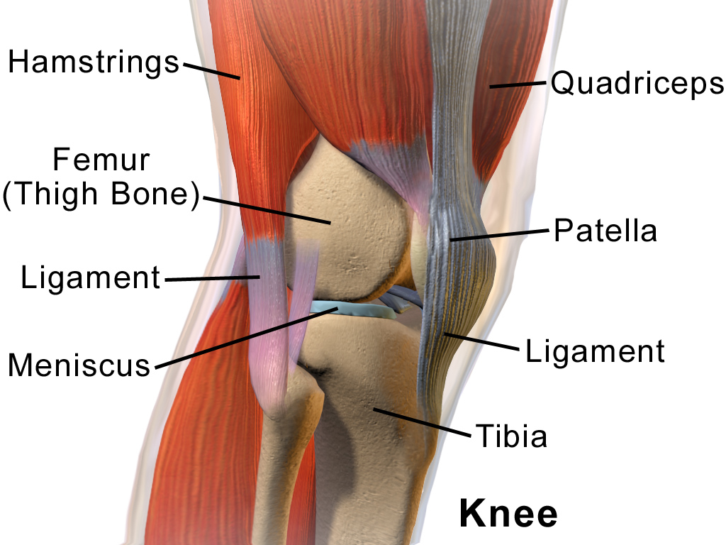
Knee
The Knee is a hinge joint that uses ligaments for stability, a Meniscus to aid in shock absorption and muscles for movement.
Meniscus
What is your meniscus?
Your Meniscus is located between your Tibia (Shin) and Femur (Thigh) bone in your knee. It comprises of 2 horse shoe shaped pieces of fibrocartilage, that play an important role within the knee. They act as a partial shock absorber, aide in the lubrication of the knee, assist in knee stability and assist in preventing damage to the articular cartilage on the tibia and femur.
How is it injured?
It is commonly injured through excessive
rotational force through the knee, this generally occurs when the knee is planted on the ground and the knee is twisted (ie the body goes in the opposite direction).
It was very common for significant pieces of meniscus to be removed through surgery in the past, although now they try to save as much as possible by only removing what is necessary. The meniscus has varying blood supply depending on the area and thus making healing of a torn meniscus very difficult.
Ligament
There are 4 main ligaments in the Knee. They are the Anterior Cruciate Ligament (ACL), The Posterior Cruciate Ligament (PCL), the Medial Collateral Ligament (MCL) and the Lateral Collateral Ligament (LCL)
ACL
The Anterior Cruciate Ligament's role in the knee is to prevent forward translation of the tibia (lower leg) in relation to the femur (thigh bone), while also providing support in pivoting type movements.
The ACL is most commonly injured in sports where the foot is planted on the ground and the person twists or pivots off that leg, or in jumping sports when the player lands on that leg. ACL rupture is most commonly managed with surgical repair, although some people do elect to live life with an ACL deficient knee. This would be something that would need to be discussed with your orthopaedic surgeon. When the ACL is injured it is very common for the MCL and meniscus to be injured as well.
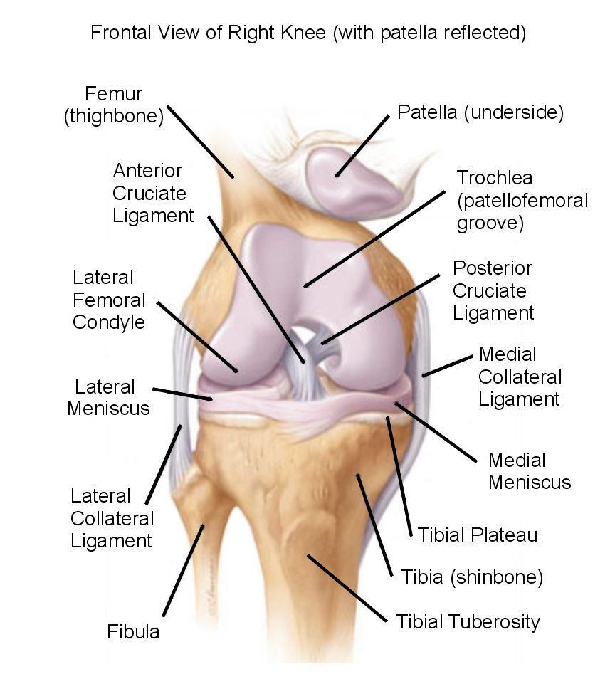
PCL
The Posterior Cruciate Ligament prevents the femur (thigh bone) from translating off your tibia. It is most commonly injured in a hyper extension of the knee or in a car accident where the knees hit the dash board.
MCL
The Medial Collateral Ligament is a strong ligament that withstands forces from the lateral (outside) of the knee to the medial (inside aspect of the knee). If the MCL is torn, depending on the severity it is most commonly managed in a specific brace. Your Physiotherapist can fit these braces.
LCL
The Lateral Collateral Ligament does the same as the MCL except that it is on the lateral aspect of the knee and resists forces from the medial to lateral aspect of the knee.
Patelofemoral Pain (PFP)
Is described as pain around the anterior to medial aspect of the knee that is caused by the articulation of the patella (knee cap) on the femur (thigh bone). If the patella is not sliding and gliding correctly over the femur this can cause knee pain. A temporary short term way to reduce pain can be through a specific taping technique, while in the long term specific strengthening is necessary.
References:
Bruckner, P & Khan, K 2006, Clinical Sports Medicine, 3rd edn, McGraw-Hill, North Rhyde, NSW.