Imaging
Medical Imaging can play an important part of confirming or excluding a specific diagnosis, although it is important to note that they should be used in conjunction with a thorough history taking and physical examination. Physiotherapists are able to refer for certain types of imaging but all types and will only refer if it is indicated.
Types of Imaging
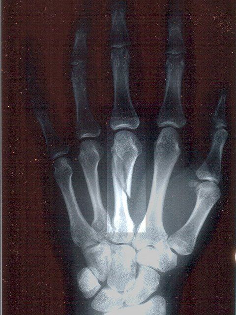
X-ray
Even though there are more detailed scans available today, the plain film x-ray still plays an important part in investigating certain conditions. X-rays can detect abnormalities in relation to bones. Specifically related to sports injuries they can detect dislocations, fractures, calcium deposits, bone spurs and leg length discrepancies. It is important to note that it is common for them to not detect stress fractures.
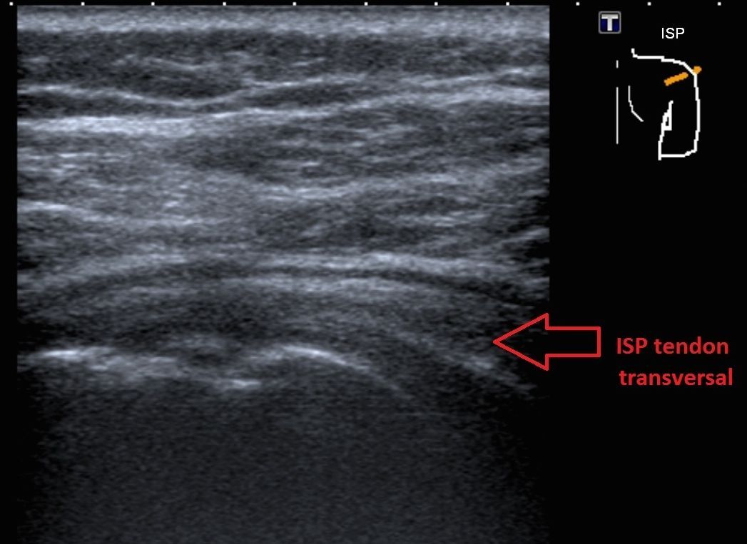
Diagnostic Ultrasound
Is a radiation and pain free technique of visualising soft tissue structures within the body. In sports injuries it is commonly used to scan shoulder tendons, Achilles tendon, ankle ligaments, hernias, muscle tears and contusions. The disadvantages of ultrasound is that it can not scan deeper structures, for example: the meniscus in the knee, the images are not as detailed as compared to an MRI.
MRI
Is a large magnet spinning around the body, realigning certain molecules that then produces an image, it is non invasive but can be a bit noisy and does not use the radiation technique that x-ray and CT uses. The clarity of the scans produced is very detailed in relation to soft tissue structures and can pick some smaller boney abnormalities that x-ray cannot. Although usually CT is the preferred choice of scan when looking for highly detailed scans of the bones.

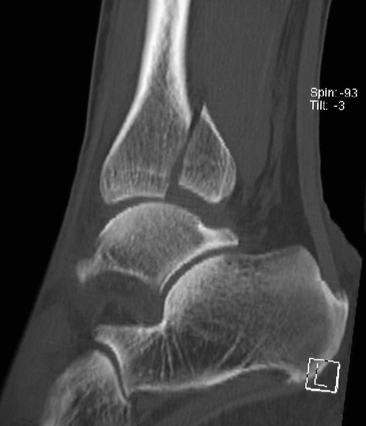
CT Scan
Is a very detailed 2D x-ray scan that can then be used to stitch together and make a 3D image of certain structures. It is a very good scan for detecting fractures, stress fractures in smaller bones of the body and the spine. One big disadvantage of CT scanning is the large amount of radiation that is emitted through the body.
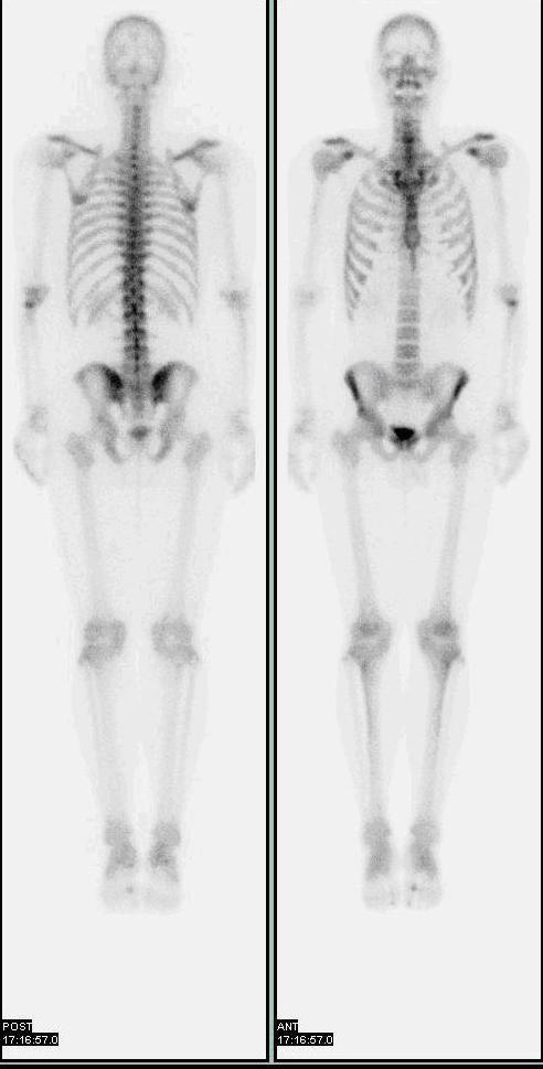
Bone Scan
Bone scans utilise nuclear medicine to reveal information through gamma ray emission. A radiopharmacutical is injected into the blood stream and after a set period of time, the person is then scanned and the machine detects areas of the body where there is increased blood flow and bone turn over. This could be areas of inflammation, infection, fracture and bone lesions (tumors). In sports medicine these scans are useful in relation to detecting stress fractures.
References
Bruckner, P & Khan, K 2006, Clinical Sports Medicine, 3rd edn, McGraw-Hill, North Ryde, NSW.